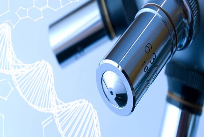
European researchers, surgeons and research institutes have just taken up a new microtechnical challenge in the service of health, with the launch of the first European prototype of an endoscopic robot for laser microsurgery of the vocal cords, which has just successfully passed pre-clinical tests.
Vocal cord surgery concerns cancerous lesions, mainly tobacco-related (3,000 new cases per year) but also, increasingly, benign lesions in people who demand a lot from their voices. In order to guarantee the preservation of the patient's voice, it requires a precision of around 50 to 100 micrometers (about the diameter of a hair). At present, however, surgery uses a laser whose source is located 40 centimetres from the patient's mouth, which limits its manoeuvrability and the precision of the gesture. Moreover, the surgeon operates through a microscope, and the tissue to be treated must therefore be in the field of vision, which implies a very uncomfortable position for the patient, a source of post-operative cervical pain.

 To overcome these drawbacks, the European project μRALP (1)The Italian Institute of Technologies, supported by the Italian Institute of Technologies, and associating among others the FEMTO-ST Institute and the CHRU of Besançon, is developing a robot dedicated to assisted surgery of the vocal cords. This new device consists of a flexible endoscope that allows visualization of the vocal cords and brings the laser source inside the patient, 20 mm from its target.
A real microtechnical challenge, the proposed endoscope includes cold light lighting, two miniature cameras to ensure 3D vision, a surgical laser coupled with a laser that acts as a pointer for the surgeon, and finally, the 1 cm3 micro robot that will, thanks to the images provided by the cameras, guide these lasers along the reference trajectory drawn by the surgeon directly in the image using a touch-sensitive tablet. Thanks to a fluorescence technique, filters will help to determine if there is a cancerous area at the lesion and will increase the precision of the gesture. With this new procedure, the surgeon should achieve an accuracy of about 100 micrometers.
After numerous technical research and development steps, pre-clinical tests are carried out on cadavers at the Institute of Anatomy of the University of Franche-Comté for the validation of the concept of the current prototype. Today's tests focus on endoscope insertion, 3D visualization, workstation ergonomics and laser guidance.
The characteristics of the endoscopic robot
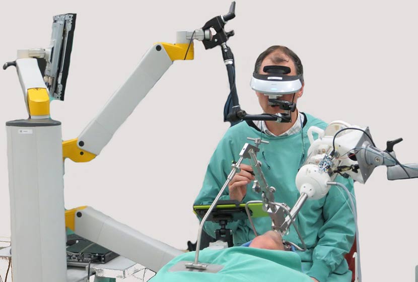
At present, vocal cord surgery suffers from several disadvantages, both for the surgeon and the patient. To improve ergonomics, the European µRALP project teams have designed a device to bring both imaging technologies and surgical tools as close as possible to the site of surgery, in the patient's body. The prototype designed is a robotic endoscope including a lighting system, cameras, and lasers, which can be remotely manipulated by the surgeon, and which also has an augmented reality visualization interface.
Access to the organs to be operated on: the robot is carried by an endoscope, a flexible rod inserted at the back of the patient's throat, as close as possible to the vocal cords (20 mm). This device can be adapted for microsurgery on other parts of the body, such as the digestive tract.
A micro-robot to finely control the laser: a micro-robot of less than 1 cm3, guided by visual feedback, ensures the orientation of the laser beam for precise surgery of the vocal cords. Concretely, the surgeon draws, on a touch-sensitive tablet displaying the preoperative images, the trajectory that the laser must adopt. By comparing the filmed images in real time with the path defined by the surgeon, the micro-robot adjusts the position and orientation of the laser beam.
A man-machine interface powered by augmented reality
Fluorescence imaging
The project developed a fluorescence imaging system to visualize cancerous tissue. Tumour cells contain specific proteins that emit fluorescent light when illuminated by a laser at a wavelength of 405 nm. Since fluorescence disappears rapidly after death, this property is not tested in cadaver tests. However, scientists are testing the device on vocal cord tumours just after they are removed from the CHRU. They exploit the properties of fluorescence on different spectral bands to help with diagnosis, the "gold standard" of which today remains histological examination.
Augmented reality visualization
In the augmented reality helmet will eventually be projected the pre-operative images, the images filmed in real time by the cameras, as well as the fluorescence signal. This device will make it possible to completely remove the tumor, minimizing damage to healthy tissue.
An intelligent and safe system
The micro-robot implements adaptive control algorithms. Ultimately, it should be able to adapt to the changing appearance of the surgical site during the procedure.
The system also improves the safety of the operation, for example by filtering out any possible tremors of the surgeon.

Complementary partners
This collaborative European µRALP project brings together five institutions from three countries. Led by the Italian Institute of Technology (IIT), it associates the FEMTO-ST Institute (UFC/CNRS/ENSMM/UTBM), the Universities of Hanover (Germany) and Genoa (Italy) and the CHRU of Besançon.
 The Istituto Italiano di Tecnologia (IIT)The project coordinator, Leonardo de Mattos, is responsible for the overall management of the project, dissemination of knowledge and exploitation of results.
IIT has been involved in the development of remote operation and automation systems for microsurgery, integrating the different software subsystems through a simple and intuitive user interface.
The IIT's contribution was very important throughout the experiments and tests, in particular for its prior technical expertise on the motorized handling of the surgical laser, as well as for its work on the ergonomics and safety of the system.
The FEMTO-ST Institute was involved in the project through two teams: the "MiNaRoB" (MicroNanoRobotics Biomedical) team, led by Nicolas Andreff (University of Franche-Comté), from the "AS2M" department (Automation and Micro-Mechatronic Systems), designed several innovative solutions for the laser micromanipulator, created a high-frequency imaging device and developed original control algorithms. This team has also played a key transversal role, participating in endoscope design, software development and augmented reality, bringing its expertise in the fields of mechatronic and optomechatronic micro-systems, visual servoing (visual feedback guidance), multi-view geometry and rapid imaging systems.
The "PIM" team (Photonics for Medical Instrumentation), directed by Bruno Wacogne (CNRS), from the Optics Department, contributed to the project for optical instrumentation and optical characterization of biological tissues by fluorescence.
The German partner "Leibniz Universität Hannover". (LUH), represented by Tobias Ortmaier and his team "Institute for Mechatronic Systems", with a strong experience in projects linking engineering and surgery, has made a major contribution to the technological developments of the flexible endoscope body, the hardware integration (including a surgical fiber laser), the monitoring of tissue deformations and augmented reality.
At the CHRU of Besançon, the ENT departmentThe clinical investigation centre (CIC) Inserm 1431 (Lionel Pazart, Bruno Wacogne), led by Laurent Tavernier, and the clinical investigation centre (CIC) Inserm 1431 (Lionel Pazart, Bruno Wacogne) took part, with their Italian counterparts, in the definition of the medical, surgical, clinical and regulatory specifications and constraints relating to the µRALP project.
A multimedia database including clinical information and images has been created, thus helping to gather and share knowledge and data on the relevant pathologies.
Teams from the Besançon University Teaching Hospital have also set up an ex vivo clinical trial in order to assess the relevance of using the optical fluorescence properties of biological samples for the differential diagnosis of pathological lesions; the anatomopathology department, represented by its head of department Séverine Valmary-Degano, has also provided assistance for the histological analysis of samples.
Finally, the ICC and the ENT servicein collaboration with the anatomy laboratory of the University of Franche-Comté directed by Laurent Tatu, coordinated the pre-clinical tests on cadavers, with the aim of validating the concept of the robotic endoscope. The cadavers used were those of subjects who had donated their bodies to Science according to the legislation currently in force. The trials were conducted in accordance with the usual ethical rules governing this type of scientific procedure.
The "Università degli Studi di Genova" (UNIGE) and its ENT department, headed by Giorgio Peretti, participated in the definition of specifications, medical constraints and surgical risks, bringing its extensive expertise on the medical protocols involved to guide the technological developments within the μRALP project.
THE UNIGE has also provided numerous iconographies of laryngeal pathologies, laser microphonosurgery (voice microsurgery), and laser-tissue interaction, to feed and enrich the online database.
Prof. Giorgio Peretti and his team also provided valuable assistance during discussions, project orientation and testing, thanks to their experience in otorhinolaryngology but also in the field of technological innovations adapted to biomedical applications.
Project Financing
μRALP is a three-year "STREP" project (January 2012 to March 2015) funded by the European Commission to the tune of 2.65 million euros. The funds were obtained under the call of the 7th Framework Programme for Research and Development (Framework Programme 7) on Information and Communication Technologies (ICT) for "cognitive and robotic systems operating in real-world environments".
Eventually, this medical device will bring real added value to phonosurgery (voice surgery). In addition, the devices designed as part of this project will be able to be used in other surgical robotic systems. For example, the FEMTO-ST Institute and the CHRU are already working on the design of an endoscope for laser microsurgery of the digestive tract.
(1) μRALP (pronounced "Microralp"): microtechnologies and systems for robot-assisted laser phonosurgery (phonosurgery laser-assisted microrobotic laser)
Photos: Surgeon-robot interface developed in the µRALP project by the Istituto Italiano di Tecnologia and Leibniz Universität Hannover.
© L. Godard/UFC